
Lesions that resemble acne
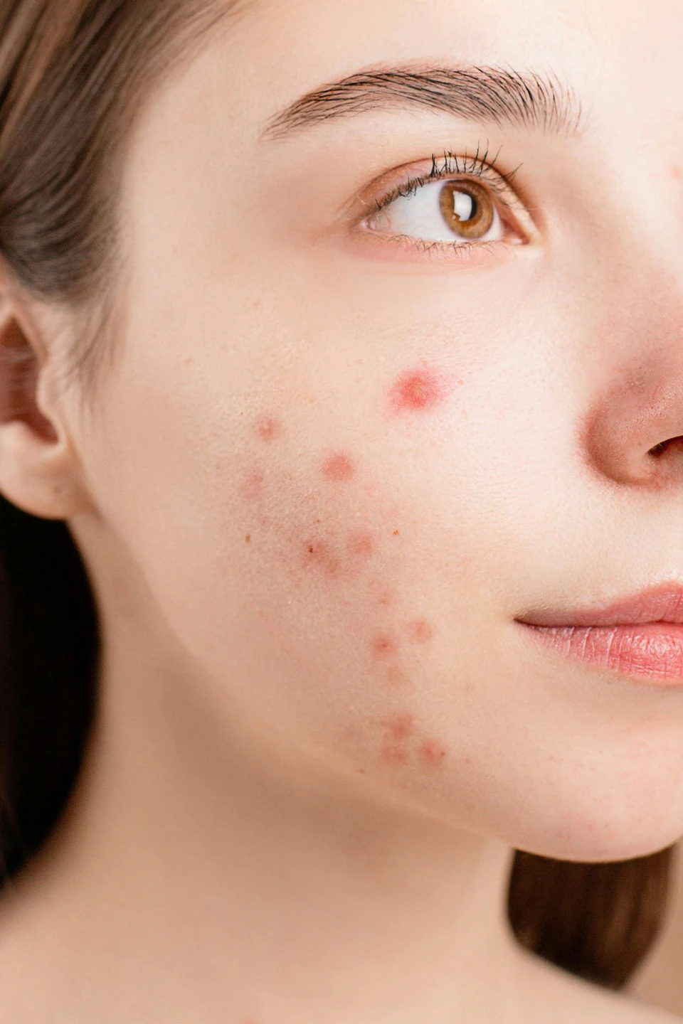
Some facial skin lesions can look like acne at first glance.
Sebaceous gland hyperplasia
This commonly occurs on the face, especially the forehead, in older adults. Lesions are small yellow bumps with a central depression.
Syringoma
This is a common sweat-duct growth. Small bumps may be skin-colored, pale yellow, brown or pink and measure about 2–3 mm. They are scattered around the eyes, especially the lower eyelids, and on the cheeks. The Thai term sometimes used is “stone acne.”
Trichoepithelioma
These growths arise from hair-follicle cells. They are small, round and smooth, skin-colored or slightly red, and about 2–4 mm in size. They may occur in clusters or be scattered over the face and often begin in childhood. Depending on their size and number, treatments described include electrosurgery, laser, liquid nitrogen, dermabrasion or excision.
Adenoma sebaceum
These growths appear as shiny, reddish-brown bumps. Close inspection may reveal numerous tiny blood vessels. They usually occur beside the nose, on the cheeks and chin, and may also appear on the forehead, scalp and ears.
They can occur with tuberous sclerosis, which may include thickened collagen tissue called a shagreen patch, leaf-shaped pale patches called ash-leaf spots, calcium deposits in the brain, growths in the eyes and kidneys, and abnormalities of bones and teeth. Pulmonary lymphangiomyoma may occur in women of reproductive age.
The condition has autosomal dominant inheritance. Reports describe intellectual disability in more than 60–70% and seizures in 80–90% of affected patients. Brain imaging, including computed tomography, is considered according to each patient’s needs. Seizures require treatment, and family-planning advice is relevant: a child of an affected parent may have a 50% chance of inheriting the condition, regardless of the child’s sex. For cosmetic management, the bumps may be removed with electrosurgery, laser or dermabrasion.
Microscopic differences
These acne-like lesions have different histopathological appearances. Syringomas form small ducts in the upper dermis, lined by two layers of epithelial cells, with a tadpole-like shape. Trichoepitheliomas contain groups of basaloid cells and keratin-filled horn cysts. “Adenoma sebaceum” is a misnomer: these lesions are actually angiofibromas.
Article images
References
The information on this website is for educational purposes only and is not a substitute for diagnosis or treatment by a physician. If you have symptoms or questions about a skin condition, please consult a dermatologist. Contact Asoke Skin Hospital at asokeskin.com
Medical disclaimer